Green tales: Vivid stories shared by People's Daily representative at Global Media Congress embody China's practice of ecological civilization construction
Editor's Note:
Under the guidance of Chinese President Xi Jinping, China has seen historic changes in ecological conservation, upholding the belief that lucid waters and lush mountains are invaluable assets, a core concept of Xi's Thought on Ecological Civilization.
In the practice of ecological civilization construction, there are numerous vivid examples, whether in the battles for blue skies, clean waters, and green forests, as well as wildlife protection. Many touching stories have emerged from these practices.
On November 14, at the Global Media Congress held in Abu Dhabi, the UAE, a representative from the People's Daily shared the newspaper's reports on ecological civilization, which sparked enthusiastic responses from the audience. Particularly attention-grabbing were three stories, which not only deeply impressed the audience with informative details and poetic writing, but also showcased a microcosm of China's construction of ecological civilization, as well as the power of the media in the process.
The three stories narrated how a village achieved a win-win outcome for both the economy and the environment through ecological protection and sustainable development, underscored the necessity of every individual becoming a protector, builder, and beneficiary of the environment, and demonstrated how systematic and scientific environmental protection work and media promotion can lead to significant transformations.
In recent years, the People's Daily has been committed to telling China's ecological stories and showcasing the achievements of China's ecological civilization construction. As efforts to strengthen ecological civilization construction continue, and as progress is steadily made toward achieving dual carbon goals, more such ecological cases are expected to emerge. These cases will become vibrant stories in the new era, underlining the country's commitment and advancements in environmental sustainability.
First Story: A village's transformation
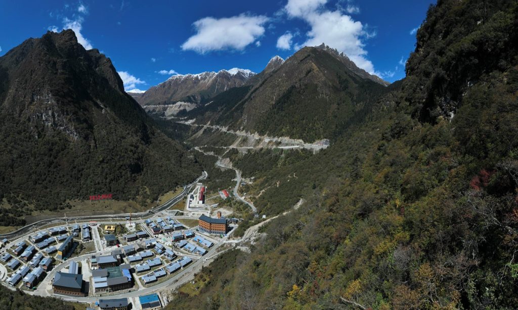
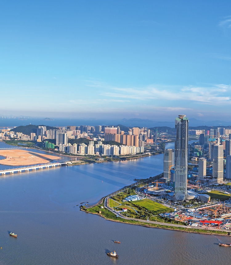
Yucun village is located in Anji county of East China's Zhejiang Province. The village was plagued by pollution and safety issues more than 20 years ago, as people there blew up mountains for mining purposes and built cement factories, which filled the sky with smoke and dust, and made streams cloudy.
In 2003, Xi Jinping, who was the then secretary of the Zhejiang Provincial Committee of the Communist Party of China (CPC), launched the "Thousand Villages Demonstration and Ten Thousand Villages Renovation" project for better green development. After the implementation of this project, Yucun village made up its mind to shut down its polluted mines and cement factories.
How could Yucun village then be further developed? In August 2005, Xi proposed that "clear waters and lush mountains are invaluable assets," during a visit to Yucun. Yucun implemented this concept and started making efforts in greening and beautifying the village. It gradually developed into a tourist destination with beautiful scenery all year round, and was included in a list of 44 villages from 32 countries termed the "Best Tourism Villages" by the United Nations World Tourism Organization (UNWTO).
From "selling stones (mineral products)" to "selling scenery," the clear waters and verdant mountains have become a source of income for the villagers of Yucun. Yucun village received 700,000 tourists in 2022, and the per capita income of the local villagers reached 64,000 yuan ($8,780).
Fifteen years later, President Xi revisited Yucun village in March 2020 during an inspection trip to Zhejiang. Seeing the changes there along the way, he said that the development of Yucun proves that green development is the right path, which should be carried on into the future.
Great changes have taken place in many villages in China including Yucun. The "Thousand Villages Demonstration and Ten Thousand Villages Renovation" project, which has resulted in thousands of beautiful villages and benefited lots of Chinese farmers through its green development concept, was awarded the Champion of the Earth award, the UN's highest environmental honor.
"Clear waters and lush mountains are invaluable assets" has become a broad consensus in today's China. In the new era, China has accelerated the transformation of its development mode into a green and low-carbon one, optimizing its industrial and energy structures, and advocated a green and low-carbon lifestyle. Today, China ranks first in the world in the scale of renewable energy development and utilization, and in the production and sales of new energy vehicles.
The story of Yuncun village demonstrates that to protect the environment is to protect productive forces, and to improve the environment is to develop productive forces, said the People's Daily representative.
To promote ecological civilization, we must scientifically grasp the dialectical unity between development and protection, and firmly establish and practice the concept that "clear waters and lush mountains are invaluable assets," said the representative.
Also, to do a good job in covering China's ecological civilization, Chinese journalists must adhere to Xi Jinping's Thought on Ecological Civilization as a guide and stand from the height of harmonious coexistence between man and nature, the representative added.
Second Story: Blue sky diary
Wang Ruchun, a 78-year-old retired worker in Shijiazhuang, North China's Hebei Province, is a shutterbug. Wang had been photographing the same piece of sky every morning since New Year's Day in 2014.
But why is he so obsessed with photographing the sky? In the earlier years, some areas in northern China were frequently plagued by large-scale and severe haze in autumn and winter, and the fine particulate matter (PM 2.5) became a major concern for people.
"At that time, I rarely captured the blue skies and white clouds in my camera," Wang recalled. "After the Chinese government launched the Three-Year Action Plan for Winning the Blue Sky Defense Battle, there were more and more blue skies."
A set of data echoed Wang's "sky dairy." The number of good air quality days in 2013 was only 43, but it increased to 234 in 2022.
People's Daily reporters covered and followed up on Wang's story, a vivid example showcasing the continuous improvement of China's air quality and environment. On June 5, 2022, the World Environment Day that year, the People's Daily released a multimedia story titled "Sky Diary: 3,000 photos record improvements to air quality in northern Chinese city of Shijiazhuang." The story became a hot search topic on social media, with many of Chinese netizens sharing photos of the blue skies of their hometowns.
In a response to netizens' demands, the People's Daily and Chinese Ministry of Ecology and Environment jointly launched a photography activity that invited Chinese netizens to take more photos of the blue skies of their hometowns. The activity received more than 300 million views.
As a netizen commented, where there is effort, there will be returns. Blue skies and white clouds don't lie. A set of convincing data released by the Ministry of Ecology and Environment showed that China has become the fastest country in the world to improve air quality. In 2022, 86.5 percent of the days in Chinese cities at prefecture level and above registered good air quality, and the number of days with heavy pollution fell to less than 1 percent for the first time.
In China, "the environment is the people's livelihood, the greenery of the mountains is beauty, and the blue sky is happiness" has become a societal consensus.
The sky diary story affirms that a good ecological environment is indispensable to the conservation of people's livelihoods. The construction of ecological civilization can best bring a sense of direct benefit to the people, and when the environment is improved, people have a more profound lived experience.
It's necessary to let everyone become the protector, builder, and beneficiary of the ecological environment in the process of promoting ecological civilization, said the People's Daily representative.
To do a good job in ecological civilization reporting, Chinese journalists must tell the stories of people excellently and share the sense of direct benefit among ordinary people, so that the broader audience can resonate, the representative noted.
Third Story: A sea of forests
The story of Saihanba is now well known throughout China, but once it was unfamiliar to most. This inspiring tale of decades of silent toil and guardianship over a forest became known to the world through a series of reports by the People's Daily.
The Saihanba Mechanized Forest Farm, located more than 400 kilometers from China's capital Beijing in the northernmost part of Hebei Province, was established in 1962. At that time, it was a desert wasteland, where "yellow sand hid the sun, and birds had no trees to perch on," with the average annual snow cover lasting seven months.
Chen Yanxian, now an octogenarian, represents the first generation of foresters. She recalled: "When we first arrived in Saihanba, we ate dark bread and drank water from melted snow, and I cannot count the number of difficulties we overcame."
Year after year, with generation after generation of steadfast commitment, Saihanba eventually became a man-made forest spanning a million acres, the largest of its kind in the world, creating a miraculous transformation from barren land to lush forest.
However, not many were aware of the touching story of the Saihanba farm. Upon discovering this lead, the People's Daily dispatched journalists for in-depth exploration and focused reporting.
From frontpage stories to key commentaries, from visual specials to integrated media products, and even a published book titled Beautiful Saihanba, the People's Daily told the remarkable story of Saihanba from all angles and perspectives.
On August 4, 2017, the newspaper published a front-page headline article "Saihanba: An Example of Ecological Civilization Construction," which stated, "Plants and trees do not speak, but walking in Saihanba, every blade of grass and tree reminds us: There is no substitute for the ecological environment, unnoticed when present, but sorely missed when lost… Mountains and rivers do not talk, but walking in Saihanba, every hill and stream tells us: Lucid waters and lush mountains are invaluable assets; they are gold and silver mountains in themselves."
An editorial published that same day mentioned that the half-century of glorious achievements of Saihanba is a vivid microcosm of green development and a classic example of ecological civilization construction.
The commentator's observation on that day also concluded that "time will not fail anyone. The story of Saihanba tells us that as long as we can sow the seeds and see the roots, we can ultimately build firm ecological barriers and write green legends."
Today, the miracle created by the people of Saihanba is widely celebrated, having received the United Nations' highest environmental honor, the "Champion of the Earth" award in 2017.
In August 2021, President Xi went on an inspection tour of Saihanba. Speaking with staff representatives of the farm, Xi praised generations of workers there for embodying the Saihanba Spirit through concrete actions. The Saihanba Spirit emphasizes staying true to the original aspiration, being hardworking and enterprising, and pursuing green development.
The story of Saihanba is just a microcosm of China's efforts to restore its ecosystems. Over the last decade, China has planted 10.2 billion acres of trees. About one-quarter of the global increase in green area since the beginning of this century comes from China.
The Saihanba story teaches us that advancing ecological civilization construction depends not only on material strength but also on spiritual power. To properly report on ecological civilization, it is essential to elucidate the scientific method of coordinating the management of mountains, rivers, forests, fields, lakes, grasslands, and sands, to apply a systematic approach, and to master the combination of integrated reporting techniques, the representative said.