Closing 'greenhouse' won't boost European EV competitiveness: Global Times editorial
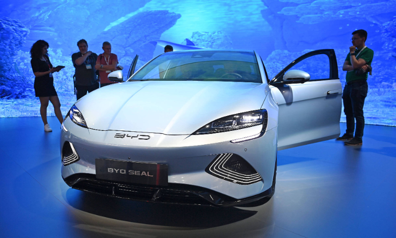
To tell the truth, when Chinese new energy vehicles shone brightly at the recent 2023 International Motor Show in Germany, we heard some envious and even jealous remarks. But we didn't expect Europe's response to be so "excessive." On September 13, Ursula von der Leyen, President of the European Commission, announced that they are launching an anti-subsidy investigation into Chinese electric vehicles (EVs). The European Union's decision is regrettable because while it acknowledges its own issues, it has chosen the wrong direction in haste and has not found the right solution to the problem.
The reasons provided by the European Union for initiating this anti-subsidy investigation are unfounded. It claimed that Chinese EVs receive "enormous state subsidies," resulting in artificially reduced prices that disrupt the European market. However, this does not align with facts. Chinese EVs are sold at significantly higher prices in Europe compared to China, whereas certain European EVs are priced lower in the Chinese market than in Europe.
Currently, Chinese EVs do not have a high market share in Europe, but they are gaining momentum. This has nothing to do with subsidies. Chinese EV companies have achieved "high quality and reasonable prices" by leveraging technological advancements and innovation, lowering costs, and improving overall quality, which has won the favor of consumers.
For both European consumers and major European car companies, Chinese EVs are not a "wolf" but a beneficial presence. EVs produced in Europe are often sold at high prices. The entry of Chinese EVs has provided European consumers with more and better cost-effective options, which is a tangible benefit. Any crackdown on Chinese EVs is bound to harm the affordability that European citizens currently enjoy.
A European Union diplomat told the media, "We cannot afford to lose our car industry." This statement unveils the true intention behind EU's actions: protectionism under the guise of "fair competition." The EU claims to "protect" Europe's automotive industry, but adopting policies of trade protectionism has been proven ineffective and costly in the past. The traditional European automotive industry has been strong and lying in its comfort zone for many years, which has led to a lack of drive for innovation in EVs and competitiveness. To change this situation, it is essential to step out of the comfort zone and enhance the competitiveness of their products in a fully competitive market.
If Europe lacks the confidence and courage to win the market through fair competition, it will be impossible to establish competitiveness in the EV industry. Keeping the EV industry in a protective green house will never lead to its growth and strength. Chinese EVs serve as a catalyst and motivation for the European EV industry to strive for innovation. Trade barriers cannot bridge the innovation gap; it will only exacerbate the situation further.
As the Chinese Ministry of Commerce responded, the automotive industries of China and Europe have formed a mutually beneficial relationship, so any harm to one side will also harm the other. The Chinese market is the largest overseas market for many EU car companies, and China provides a favorable business environment for European cars. If you take a look at the roads in Germany, you will see mostly German cars, while in France, you will see mostly French cars. The same goes for Japan and South Korea. However, on Chinese roads, you can find cars from all over the world, which vividly reflects the openness and diversity of the Chinese market. All of this should be cherished and valued by Europe.
In interpersonal relationships, reciprocity is important. China and Europe should create a fair, non-discriminatory, and predictable market environment for the mutual development of the electric car industry. They should jointly oppose trade protectionism and work together to address global climate change and achieve carbon neutrality. Particularly, the EU itself is also a victim of protectionism. The Inflation Reduction Act enacted by the US last year used similar tactics to protect its domestic industries, which caused strong opposition in Europe, with many saying, "The Americans stabbed us in the back." Now, the EU is responding to foreign competitors with the same mind-set, and it should feel ashamed of its decision today.
In her speech on Wednesday, von der Leyen mentioned the example of the solar industry, stating that "we have not forgotten how China's unfair trade practices affected our solar industry." The solar industry is indeed a worthy example to review. In 2013, the EU followed the US in imposing anti-dumping tariffs on imported solar panels from China, citing the same reason of "unfair subsidies." However, the result was that because of lack of competition, the European solar industry languished, and many companies increased costs by importing Chinese products through other channels.
Looking back today, we can draw two lessons from what the solar industry suffered: First, competitiveness cannot be gained through protectionism, and blindly engaging in protectionism often backfires; second, trade disputes and differences ultimately need to be resolved through mutual negotiation. We hope that the EU can extract the correct information from the case of the solar industry, listen more to the voices of the business community, and have fewer politicized interpretations. After all, towering trees cannot grow in a greenhouse, and a steel-winged eagle cannot fly out of a birdcage.