Cells’ stunning complexity on display in a new online portal
Computers don’t have eyes, but they could revolutionize the way scientists visualize cells.
Researchers at the Allen Institute for Cell Science in Seattle have devised 3-D representations of cells, compiled by computers learning where thousands of real cells tuck their component parts.
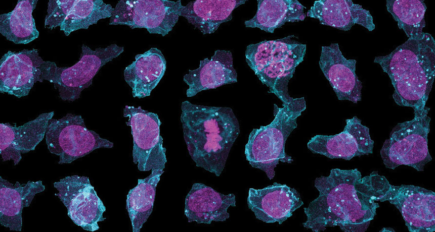
Most drawings of cells in textbooks come from human interpretations gleaned by looking at just a few dead cells at a time. The new Allen Cell Explorer, which premiered online April 5, presents 3-D images of genetically identical stem cells grown in lab dishes (composite, above), revealing a huge variety of structural differences.
Each cell comes from a skin cell that was reprogrammed into a stem cell. Important proteins were tagged with fluorescent molecules so researchers could keep tabs on the cell membrane, DNA-containing nucleus, energy-generating mitochondria, microtubules and other cell parts. Using the 3-D images, computer programs learned where the cellular parts are in relation to each other. From those rules, the programs can generate predictive transparent models of a cell’s structure (below). The new views, which can capture cells at different time points, may offer clues into their inner workings.
The project’s tools are available for other researchers to use on various types of cells. Insights gained from the explorations might lead to a better understanding of human development, cancer, health and diseases.
Researchers have already learned from the project that stem cells aren’t the shapeless blobs they might appear to be, says Susanne Rafelski, a quantitative cell biologist at the Allen Institute. Instead, the stem cells have a definite bottom and top, a proposed structure that’s now confirmed by the combined cell data, Rafelski says. A solid foundation of skeleton proteins forms at the bottom. The nucleus is usually found in the cell’s center. Microtubules bundle together into large fibers that tend to radiate from the top of the cell toward the bottom. During cell division, microtubules form structures called bipolar spindles that are necessary to divvy up DNA.
One surprise was that the membrane surrounding the nucleus gets ruffled, but never completely disappears, during cell division. Near the top of the cell, above the nucleus, stem cells store tubelike mitochondria much the way plumbing and electrical wires are tucked into ceilings. The tubular mitochondria were notable because some researchers thought that since stem cells don’t require much energy, the organelles might separate into small, individual units.
Old ways of observing cells were like trying to get to know a city by looking at a map, Rafelski says. The cell explorer is more like a documentary of the lives of the citizens.